

What is Magnetic Resonance Cholangiopancreatography (MRCP)?
Introduction:
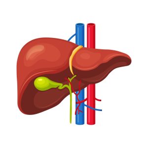
Magnetic Resonance Cholangiopancreatography (MRCP) procedure is a specialized, non-invasive imaging technique used to evaluate the biliary and pancreatic ductal systems. It is a type of Magnetic Resonance Imaging (MRI) that provides detailed, high-resolution images of the liver, gallbladder, bile ducts, pancreas, and pancreatic ducts without requiring invasive procedures such as endoscopic retrograde cholangiopancreatography (ERCP).
MRCP abdomen plays a crucial role in diagnosing various hepatobiliary and pancreatic diseases, including gallstones, bile duct strictures, pancreatic cysts, and tumours. This imaging modality has significantly improved patient care by reducing the need for diagnostic endoscopic procedures and avoiding their associated risks.
How Does MRCP Work?
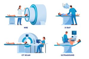
An MRCP abdomen utilizes magnetic resonance imaging (MRI) technology to generate detailed images of the bile ducts, gallbladder, liver, pancreas, and pancreatic ducts. Unlike traditional imaging techniques such as X-rays or CT scans, MRCP does not use ionizing radiation. Instead, it relies on strong magnetic fields and radio waves to create high-contrast images of soft tissues and fluid-filled structures.
Basic Principles of the MRCP Procedure:
- Magnetic Field: The patient is placed inside an MRI scanner, which generates a strong magnetic field. This aligns hydrogen protons in the body.
- Radiofrequency Pulses: The machine emits radio waves, which temporarily disturb the alignment of hydrogen protons in the tissues.
- Signal Detection: When the radio waves are turned off, the protons return to their original alignment, releasing energy captured by detectors.
- Image Formation: A computer processes the detected signals to create detailed images of internal organs, particularly highlighting the biliary and pancreatic ducts due to their fluid content.
The MRCP images are typically obtained using heavily T2-weighted MRI sequences, which enhance the visibility of fluid-filled structures while suppressing surrounding solid tissues. This makes the bile and pancreatic ducts appear bright and well-defined, facilitating accurate diagnosis.
Why do I need an MRCP scan?
The MRCP procedure is primarily used to evaluate conditions affecting the bile ducts, gallbladder, and pancreas. Some of the most common indications for the MRCP test include:
1. Biliary System Disorders:
- Gallstones (Cholelithiasis): The MRCP procedure is highly effective in detecting stones within the bile ducts that may cause obstruction.
- Biliary Strictures: Narrowing of the bile ducts due to inflammation, scarring, or malignancies can be assessed using MRCP.
- Primary Sclerosing Cholangitis (PSC): A chronic liver disease leading to bile duct strictures, which can be visualized on MRCP.
- Biliary Atresia: A congenital condition in newborns causing bile duct obstruction.
2. Pancreatic Disorders:
- Chronic and Acute Pancreatitis: MRCP abdomen helps identify inflammation, calcifications, and structural changes in the pancreas.
- Pancreatic Duct Abnormalities: Dilation or strictures in the pancreatic duct can be evaluated to determine underlying pathology.
- Pancreatic Cysts and Tumours: The MRCP test procedure aids in distinguishing between benign cysts and malignant lesions.
3. Post-Surgical Evaluation:
- Biliary Leak Detection: After gallbladder removal (cholecystectomy) or liver surgery, an MRCP scan can help detect bile leaks.
- Post-Transplant Assessment: Liver transplant recipients may undergo MRCP to check for bile duct complications.
4. Congenital Abnormalities:
- Choledochal Cysts: Congenital dilations of the bile duct system can be effectively diagnosed with MRCP.
- Pancreas divisum: A congenital anomaly in which the pancreatic ducts fail to fuse properly, leading to ductal obstruction and pancreatitis.
Advantages of MRCP Over Other Imaging Techniques
The MRCP procedure offers several benefits compared to alternative diagnostic methods, making it the preferred choice for evaluating hepatobiliary and pancreatic diseases.
1. Non-Invasive and Radiation-Free
Unlike ERCP, the MRCP machine does not require an endoscope or the injection of contrast dye into the bile ducts, reducing the risk of complications such as infection or pancreatitis. Additionally, it does not expose patients to ionizing radiation, making it a safer option, especially for children and pregnant women.
2. High Sensitivity for Fluid-Filled Structures
MRCP abdomen provides superior visualization of bile and pancreatic ducts due to its high-contrast resolution for fluid-filled structures. It allows clear detection of obstructions, strictures, and abnormalities.
3. No Need for Contrast Dye in Most Cases
While some MRI scans require gadolinium-based contrast agents, MRCP is often performed without contrast, reducing the risk of allergic reactions or nephrotoxicity in patients with kidney disease.
4. Useful for Surgical Planning
MRCP tests help surgeons plan interventions by providing detailed anatomical information before procedures like bile duct surgery or pancreatic resections.
5. Reduces the Need for ERCP
Since MRCP imaging provides detailed images of the bile and pancreatic ducts, many patients can be diagnosed without undergoing invasive procedures. ERCP is now primarily used for therapeutic interventions rather than diagnostics.
Limitations and Risks of the MRCP Test
While the MRCP machine is a powerful diagnostic tool, it has some limitations:
1. Limited for Small Stones or Early-Stage Disease
MRCP abdomen may not detect very small gallstones or early bile duct cancers as effectively as ERCP. In cases where a definitive diagnosis is needed, additional testing may be required.
2. Motion Artifacts
Patients need to remain still during the scan. Movement can cause blurry images, affecting diagnostic accuracy. In some cases, breath-holding sequences are used to improve image quality.
3. Not Suitable for Patients with Certain Implants
Patients with pacemakers, metal implants, or certain aneurysm clips may not be able to undergo MRCP due to the strong magnetic field of the MRI machine.
4. Longer Scan Time Compared to CT Scans
MRCP imaging typically takes 30-60 minutes, whereas a CT scan can be completed in just a few minutes. This might be challenging for claustrophobic patients.
Preparation for MRCP procedure:
Proper preparation is essential to ensure high-quality images.
Before the Scan:
- Patients are advised to fast for 4-6 hours before the procedure to reduce the presence of fluid and air in the stomach and intestines, which can interfere with imaging.
- Metallic objects, including jewellery, watches, and hairpins, must be removed.
- Patients with claustrophobia should inform their doctor, as mild sedation may be needed.
During the Scan:
- The patient lies on a movable MRI table that slides into the scanner.
- A coil (receiver device) is placed around the abdomen to capture detailed images.
- The patient may be asked to hold their breath briefly during imaging sequences.
After the Scan:
- No recovery time is required unless sedation is used.
- A radiologist analyzes the images and provides a report to the referring physician.
Difference Between ERCP and MRCP
Both ERCP (Endoscopic Retrograde Cholangiopancreatography) and MRCP (Magnetic Resonance Cholangiopancreatography) are used to evaluate the bile ducts, pancreas, and gallbladder, but they differ in technique and purpose.
- ERCP: A minimally invasive endoscopic procedure that uses a flexible tube with a camera to examine and treat bile or pancreatic duct blockages (e.g., stone removal, stent placement).
- MRCP: A non-invasive MRI-based imaging technique used to diagnose conditions like bile duct stones, strictures, or tumours without the need for an endoscope.
Key Difference: ERCP is both diagnostic and therapeutic, whereas MRCP is purely diagnostic and avoids the risks of an invasive procedure.
Conclusion:
Magnetic Resonance Cholangiopancreatography (MRCP) scan is a highly effective and non-invasive imaging technique for diagnosing disorders of the bile ducts, gallbladder, and pancreas. It provides high-resolution images without the risks associated with invasive procedures like ERCP.
MRCP imaging is particularly useful in detecting gallstones, bile duct strictures, pancreatic abnormalities, and post-surgical complications. Its ability to offer detailed, radiation-free imaging makes it a preferred choice for both doctors and patients.
While the MRCP test has some limitations, its advantages far outweigh them, making it a valuable tool in modern medical diagnostics. If your doctor has recommended an MRCP scan, understanding its purpose and process can help ease any concerns and ensure a smooth experience.
Always consult your healthcare provider for further consultation to determine if MRCP is the right diagnostic tool for your condition.